Late-phase clinical trials have evolved. They’re no longer just about large sample sizes and long timelines—they’re about data depth, real-world insights, and precision.
And among all the growing data points in these trials, medical imaging has emerged as one of the most complex—and critical—to manage.
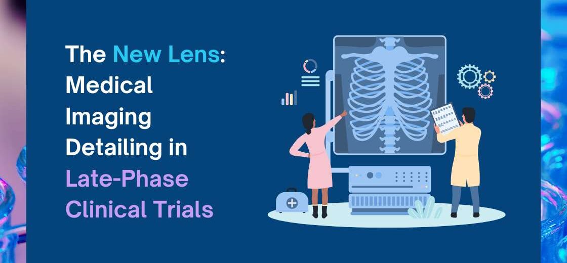
In oncology and other image-intensive therapeutic areas, imaging is no longer just part of the dataset. It is often the deciding factor. And with that, comes a new need: Imaging Detailing.
So, What Does Imaging Detailing Mean in Late-Phase Trials?
Imaging detailing isn’t just about storing scans. It’s about the systematic handling of everything that surrounds those images—from acquisition to interpretation.
It includes:
- Structured image acquisition based on protocol specifications
- Lesion-level measurement across visits
- Standardized grading workflows with adjudication
- Data quality and traceability controls
- Equipment and technician qualification tracking
- Secure transfer, encryption, and audit readiness
- Real-time visibility of imaging progress across sites
In simple words: it’s treating imaging as a controlled scientific process, not just a clinical task.
Why It Matters: Imaging Is Now a Data Source, Not Just a File
In late-phase trials, especially oncology, ophthalmology, and orthopedics, imaging serves three strategic purposes:
- Endpoints: Imaging often provides the primary or secondary endpoint—tumor size, disease progression, retinal changes, bone alignment, etc.
- Timely Treatment Decisions: Delays or errors in imaging interpretation can directly impact when a patient is dosed, escalated, or withdrawn.
- Auditability: Regulators now expect structured image workflows, traceable reads, and calibration proof—not just scan folders and reviewer notes.
That’s why imaging detailing has become a critical capability—and not every system can handle it.
The Real-World Challenges We See
From global trials we’ve supported, here are common issues that surface when imaging isn’t detailed well:
- Sites uploading wrong format or low-quality scans
- Inconsistent lesion measurements across visits or readers
- Reviewers working in silos without centralized access
- Missing technician logs or calibration records during audits
- Bandwidth challenges delaying uploads from remote sites
- No unified workflow for image read, grading, and sign-off
Each of these can lead to data queries, deviations, or even protocol failures.
Why Detailing Needs a Specialized Platform Like MI LP
That’s where a dedicated imaging module like MI LP (Medical Imaging for Late Phase) comes in—not just to digitize but to organize, control, and enhance the entire imaging journey.
Here’s how MI LP brings structure to imaging detailing:
Modality Support & Smart Upload
Handles DICOM and other formats with a smart upload process that works even on low-bandwidth sites.
Lesion Measurement & Calibration
Guides consistent lesion-level measurement, visit by visit, with built-in calibration tools.
Multi-Grader Workflows
Supports independent reviews, adjudication, and grading trails—ensuring consistency and reducing bias.
Image Encryption & Compliance
Secure transfer with audit logs, 21 CFR Part 11 compliance, and encrypted data handling.
Reviewer & Technician Management
Tracks equipment registrations, technician qualifications, and reviewer sign-offs—no guesswork during audits.
Real-Time Notifications & Dashboards
From image receipt to grading status—get visibility into imaging progress and pending reviews.
A Closer Look at MI LP’s Imaging Intelligence
MI LP isn’t just a module—it’s an imaging operations system designed with the reality of late-phase trials in mind.
- Works across oncology, retinology, and orthopedics
- Integrates with IWRS and trial platforms seamlessly
- Configurable to fit unique study imaging protocols
- Enables structured reviews with export-ready outputs
- Built for global studies, including multi-site and decentralized trials
Final Word: Imaging Isn’t an Attachment—It’s an Asset
In late-phase trials, especially as real-world relevance and regulatory expectations increase, imaging must be treated with precision and process.
Imaging detailing isn’t about adding more work—it’s about giving structure to the work you’re already doing.
And when done right, it adds real value—better data, faster insights, and more confidence at every decision point.
MI LP helps you get there—one detailed, traceable image at a time.